|
|
Medical Neuroscience
THE HUMAN HYPOTHALAMUS: Major Anatomical Features
Prepared by John I. Johnson. Ph. D., Anatomy Department and Mark W. Hodgins, Office of Academic Affairs, College of Osteopathic Medicine, Michigan State University.
Specimen courtesy of Robert C. Switzer III, Ph. D., Director of Brain Collection in the R. H. Cole Neuroscience Laboratory, University of Tennessee Medical Center at Knoxville.
Histological preparation by Shannon K. Campbell and Tracy Pesut, R. H. Cole Neuroscience Laboratory, University of Tennessee Medical Center at Knoxville.
Prepared for Web Site use by Richard W. Carlson, Okemos, Michigan.
Select a scan level and stain below to continue:
|
|
 |
| Click on a blue line to view the Cell Stain section at that level |
|
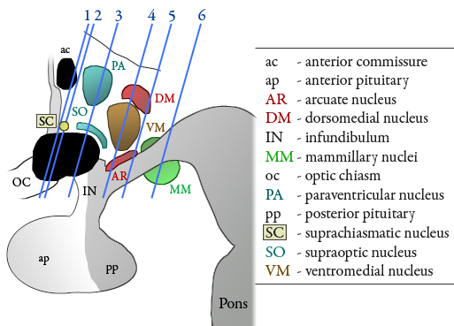
SAGITTAL OVERVIEW
This diagram represents a view of the wall of the third ventricle from the midline of the brain, and shows the six levels of the coronal sections seen in the series that follows. Dorsal is toward the top of the frame, and anterior is to the left.
At each level two sections are presented, one stained for cells by the Nissl method, and a neighboring section stained for myelinated fibers by the Weil method.
The sections are from the brain of a 21 year old male who died of traumatic injuries from an automobile accident. The locations of major hypothalamic nuclei are shown in color.
This overview diagram will be repeated to the upper left of each of the sections, and a key identifying the abbreviated labels will appear to the lower left. Outlines and labels can be seen in place on the sections, and can be removed for a better view of the neural architecture.
Hypothalamic nuclear outlines and abbreviations are color-coded to match the overview diagram. Self-testing can be done by reviewing the list of labeled structures with the labels and outlines removed from the section (find the location of each structure in the unlabeled section); turning the labels and outlines back on (by clicking on the image) serves as a key for the self-test. There is an outline drawing in the class handout for each level shown here (each pair of sections).
|
