The Sheep Brain Atlas

John I. Johnson, Keith D. Sudheimer, Kristina K. Davis, Garrett M. Kerndt, and Brian M. Winn
Radiology Department, Neuroscience Program,and Communications Technology Laboratory,
Michigan State University, East Lansing, MI

Please let us know if you are using any of these atlases or images for particular classes. This information will assist us in maintaining support for these atlases. Send this information to Josh Vincent vincen29@msu.edu

Sheep Atlas Links

External Surface Views - labeled

Superior - Lateral - Inferior


Coronal Sections
Each section is 35 micrometers thick.


NOTE: Check out the prototype glossary on section 1240.
Click on the labels to view the glossary definitions.

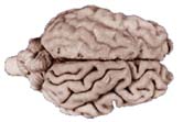
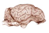
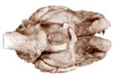
See also the Sheep Brain Dissection Guide at the University of Scranton


Supported by: Grants IBN 0131267, 0131826, and 0131028, from the National Science Foundation
Division of Integrative Biology and Neuroscience.

With much assistance from our many collaborators.

All images on this site are copyrighted and produced with the support of public funds.
Click here to learn how to use them for free