|
|
BRAIN
COLLECTION PERSONNEL
|
The
University of Wisconsin (Madison)
|
|
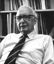
Dr.
Clinton N. Woolsey was the founder, director,
and first Chairman of the Laboratory (and then Department)
of Neurophysiology in the Medical School of the University
of Wisconsin in Madison, Wisconsin.
|
Curator
|
|
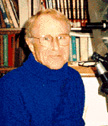
Wally
Welker
was curator of the Mammalian Brain Collection at the University
of Wisconsin. He was Professor Emeritus in the Dept. of Physiology
(formerly the Dept. of Neurophysiology) at UW in Madison,
Wisconsin. He received his Ph D in The Psychology Dept. of
the University of Chicago in 1954 (see History....);
was a Postdoctoral Fellow (NIH) at the Department
of Neurophysiology, joined the Faculty there in 1958 and
formalized the collection of mammalian brain specimens (see
History ....). |
Histologists
The
neurohistologists in the Department of Neurophysiology (now Physiology)
have played major roles in assuring the high quality of the embedding,
sectioning and staining of the brains ever since the neurohistology
lab was begun in 1950.
|
|
Helen
Brandemuehl (now deceased) established the initial
high standards for the laboratory, and trained most of the
technicians and students who have worked there. She kept
detailed protocols of procedures, routines and staining
recipes that have become standard practices for all subsequent
personnel in the Laboratory. |
|
|
Inge
Siggelkow (recently retired) was the manager and
Senior histologist of the Department of Physiology's neurohistology
laboratory. She has intimate knowledge of all procedures
used by her staff (Ms.'s Ekleberry and Meister, and with
them, carried out all the histological procedures that were
employed by her staff. She has had extensive experience
and training in neurohistological procedures, and can be
contacted by anyone who has questions about the procedures
and techniques that have been used in preparation of the
brain collections (inge@physiology.wisc.edu). Ms. Siggelkow
had access to all records of procedures used for every specimen
that exists in the Wisconsin Collection. She regularly maintained
the remaining specimens still at Wisconsin. She actively
worked with the National Museum's staff in preparing and
packing brain sections and other material that have (and
will be) transported to the National Museum. She helped
scan digital images of brain sections from specimens that
are being prepared and presented as Web-displayed brain
atlases for specimens deemed of interest to researchers,
students, and the public. |
|
|
Jo
Ann Ekleberry, Histologist, Univ. of Wis. (now retired).
Jo Ann was a devoted neurohistologist for over 26 years.
She played a major role in all aspects of histological processing
of brain specimens in our normal brain collection. She embedded
brains involving celloidin, paraffin, frozen and plastic
media. She also sectioned brains and was active in all subsequent
histological processing activities, includiong staining,
mounting sections on glass slides, cover-glassing the mounted
sections, and adjusting the saturation, color, degree of
contrast of cells and fibers to enhance visibility of neural
features. She diligently cleaned all the slide and then
organized all the slide boxes of each specimen and placed
them in metal or wooden slide boxes which were organized
all slide boxes in shelves. She also kept detailed records
of all aspects about each specimen. |
|
|
Joan
Meister, Histologist, Univ. of Wis. (now retired)
Joan was a devoted neurohistologist for over 20 years. She
played a major role in all aspects of histological processing
of brain specimens in our normal brain collection. She embedded
brains involving celloidin, paraffin, frozen and plastic
media. She also sectioned brains and was active in all subsequent
histological processing activities, including staining,
mounting sections on glass slides, cover-glassing the mounted
sections, and adjusting the saturation, color, degree of
contrast of cells and fibers to enhance visibility of neural
features. She diligently cleaned all the slide and then
organized all the slide boxes of each specimen and placed
them in metal or wooden slide boxes which were organized
all slide boxes in shelves. She also kept detailed records
of all aspects about each specimen. |
Illustrator
and Photographer
|
|
Carol
Dizack, Senior Medical/Scientific Illustrator and
Graphic Designer, Univ. of Wisconsin-Madison. Has contributed to
all aspects of preparation of this Web Site. She has arranged
and manipulated images of all brains, brain sections, as
well as composed the illustrations which are placed at the
start of each section of this electronic document, as well
as others that are presented throughout the pages of this
site. Ms. Dizack has a Masters Degree in Fine Art from the
University of Wisconsin, as well as a Degree in Commercial
Art From the Madison Area Technological College. She has
been employed at the University of Wisconsin for 40 years,
and is currently a Senior Illustrator/Designer in the department
of Media Solutions in the University of Wisconsin School
of Medicine and Public Health. She has been working with
Wally Welker, John I. Johnson and Photographer Terril P.
Stewart in preparation of illustrative materials associated
with the University of Wisconsin and the Michigan State
University Comparative Mammalian Brain Collections and for
publications and our Web Site since the beginning of her
tenure at Wisconsin. She works entirely with Macintosh computer-generated
illustrations and graphics. She is responsible for all aspects
of the illustrative composition of this Web site. She can
be reached at her e-mail address: cldizack@wisc.edu. |
|
|
Terrill
P. Stewart, Senior Photographer, Distinguished Media
Specialist, Univ. of Wisconsin. (now retired). was responsible
for all photographic activities for the Department of Neurophysiology,
as well as other Departments in the University of Wisconsin
Medical School. He took the photographs of all the brain
specimens and brain sections from the Wisconsin Comparative
Mammalian Brain Collection. All Photographs of brains presented
on this Web Site were photographed by Mr. Stewart. They
were taken by standard black and white photographic techniques
and colorized by Ms. Dizack using digital techniques. The
photographic archives of all images of animal specimens
(brain, body, and other) that comprise our brain collection
were prepared by Mr. Steward and all these will be transferred
to the National Museum when the remainder of the collection
specimens have been moved to Washington, D.C. |
Information
Technologists

Ravi
Kochhar, Information
Processing Consultant, University of Wisconsin
|

Jane Sekulski, Programmer, University of Wisconsin |

Kevin
Graeme, former Web Publisher, University of Wisconsin |

Ray
Spiess, former Web Publisher, Univ.of Wisconsin |

Mary
Walsh, Web Designer, GreenLeaf MeDia |
|

List
of Specimens
| Explore Collections | Brain
Sections | Brain Evolution |
Brain Development | Brain
Circuitry | Brain Functions
| Location and Use | Related
Web Sites | Contact Us | Search
MSU Database |
Personnel |
Home
|