
|
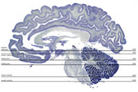
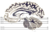
John I. Johnson, Brian M. Winn, Garrett M. Kerndt, Joseph J. Maleszewski, Myrvine Bernadotte, Prashant Vaishnava and Keith D. Sudheimer In this atlas you can view axial sections stained for cell bodies or for nerve fibers, at six rostro-caudal levels of the human brain stem. Please use the images and data from this site.
Adrianne Noe, Ph. D., Director; Archie J. Fobbs, Jr., Neuroanatomical Curator. |

All images on this site are copyrighted and produced with the support of public funds.
Click here to learn how to use them for free
Click here to learn how to use them for free