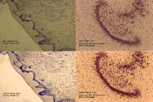
Figure 7: Photo CD vs Digital Camera
 These images illustrate the results of photographing the image on 35 mm Kodachrome film and processing through Photo CD, compared with those obtained using the Kodak DCS 200 digital camera on a Nikon Biophot microscope.
The original Photo CD image was captured by photographing on Kodachrome 35 mm film through a Zeiss GFL microscope, using a 2.5 x objective and 12.5 x ocular, using a Zeiss Ikon 35 mm camera.
The Photo CD image is from the high resolution Photo CD file (1536 x 1024 pixels, 4 Mb); that from that from the digital camera is at the original camera resolution of 220 pixels per inch (220 ppi).
All images were edited in Adobe Photoshop, and both raw and edited images were assembled in Photoshop into the composite image (200 ppi and 8 Mb) for printing.
The two sections on the left are from a horizontal section through the olfactory tubercle (ventral striatum) stained for cell bodies (Thionine stain, Nissl method). This specimen is from a Broad-footed marsupial mouse (Antechinus flavipes)
The two sections on the right are from a coronal section through the hippocampus of a Wisconsin badger (Taxidea taxus) stained for cell bodies (Thionine, Nissl method).
The images were printed on the Kodak XL 7700 dye sublimation printer, by DLM Imaging, Madison, WI.
Back to top
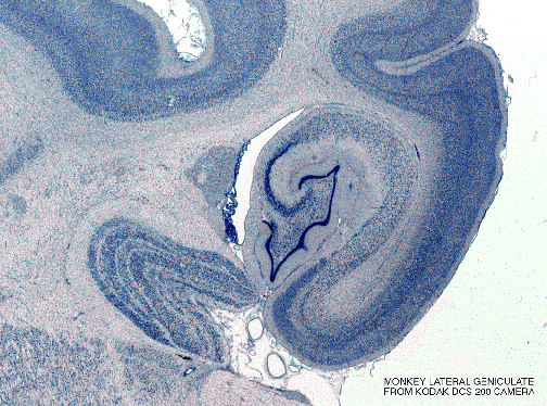
Figure 8: Digital Camera (DCS 200)
 This image was obtained using the Kodak DCS 200 digital camera with our specially designed slide holder and illuminator, and a 42 mm Leitz Summar Macro lens and extension bellows (see Methods). It was then edited in Adobe Photoshop. Excellent prints were then produced on the Kodak XL 7700 dye sublimation printer, from the original camera resolution at 220 pixels per inch (ppi), by DLM Imaging, Madison, WI.
The tissue is a coronal section through the lateral geniculate nucleus and hippocampus of a Rhesus monkey (Macaca mulatta) stained for cell bodies (Thionine stain, Nissl method).
Back to top
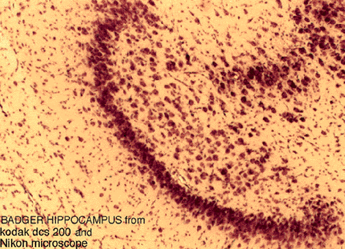
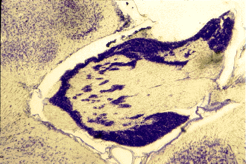
Figure 9: Digital Camera (DCS 200)
 The image was obtained using the Kodak DCS 200 digital camera on a Nikon Biophot microscope. It was edited in Adobe Photoshop, and printed from a file at the original camera resolution of 220 pixels per inch (220 ppi), on the Kodak XL 7700 dye sublimation printer, by DLM Imaging, Madison, WI.
The tissue is a coronal section through the hippocampus of a Wisconsin badger (Taxidea taxus) stained for thionine (Nissl method).
Back to top
Figure 10: Photo CD
 The original Photo CD image was captured by photographing on Kodachrome 35 mm film through a Zeiss GFL microscope, using a 2.5 x objective and 12.5 x ocular, using a Zeiss Ikon 35 mm camera. The image is from the high resolution Photo CD file (1536 x 1024 pixels, 4 Mb); it was edited in Adobe Photoshop and expanded by interpolation in Photoshop from 144 to 200 pixels per inch (ppi) and 8 Mb. for printing on the Kodak XL 7700 dye sublimation printer, by DLM Imaging, Madison, WI.
The tissue is a horizontal section through the olfactory bulb ofa Broad-footed marsupial mouse (Antechinus flavipes) stained for cell bodies (Thionine stain, Nissl method).
Back to top
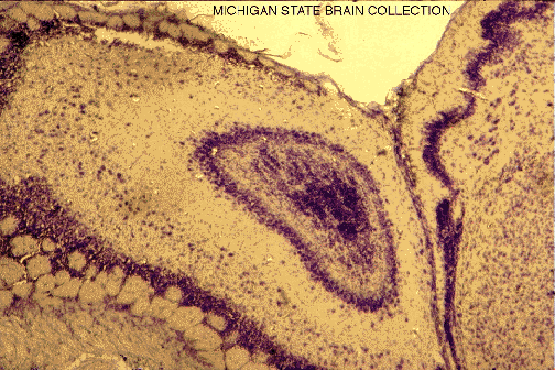
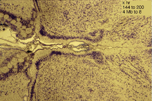
Figure 11: Photo CD
 The original Photo CD image was captured by photographing on Kodachrome 35 mm film through a Zeiss GFL microscope, using a 2.5 x objective and 12.5 x ocular, using a Zeiss Ikon 35 mm camera. The image is from the high resolution Photo CD file (1536 x 1024 pixels, 4 Mb); it was edited in Adobe Photoshop and expanded by interpolation in Photoshop from 144 to 200 pixels per inch (ppi) and 8 Mb. for printing on the Kodak XL 7700 dye sublimation printer, by DLM Imaging, Madison, WI.
The tissue is a horizontal section through the taenea tecta of a Broad-footed marsupial mouse (Antechinus flavipes) stained for cell bodies (thionine stain, Nissl method).
Back to top
Figure 12: Photo CD
 The original Photo CD image was captured by photographing on Kodachrome 35 mm film through a Zeiss GFL microscope, using a 2.5 x objective and 12.5 x ocular, using a Zeiss Ikon 35 mm camera. The image is from the high resolution Photo CD file (1536 x 1024 pixels, 4 Mb); it was edited in Adobe Photoshop and expanded by interpolation in Photoshop from 144 to 200 pixels per inch (ppi) and 8 Mb. for printing on the Kodak XL 7700 dye sublimation printer, by DLM Imaging, Madison, WI.
The tissue is a horizontal section through the trigeminal ganglion of a Broad-footed marsupial mouse (Antechinus flavipes) stained for cell bodies (thionine stain, Nissl method).
Back to top